Written by Professor Vik Veer, Consultant ENT & Sleep Surgeon · Published · Last reviewed
Quick Summary
- What it is. A persistent sensation of a lump, tightness or foreign body in the throat with no structural obstruction and no true difficulty swallowing. An estimated 45% of people experience it at some point, and it accounts for roughly 4% of ENT referrals.[1]
- First, rule out serious disease. Progressive swallowing difficulty, painful swallowing, weight loss, persistent hoarseness, a neck lump or one-sided symptoms need urgent assessment.
- Four common contributors. Laryngopharyngeal (silent) reflux, cervical inlet patch and other upper oesophageal causes, laryngeal muscle tension, and enlarged tonsils or a pharyngeal pouch. Less often, eosinophilic oesophagitis.
- Acid-blocking tablets alone do not help. A large placebo-controlled trial found no benefit of a PPI for throat symptoms at 16 weeks or 12 months.[7] An alginate is a rational, low-risk first-line option.
- What you can do now. Earlier evening meals, fewer reflux triggers, a raised bed head, left-side sleeping, weight loss and stopping smoking all have supporting evidence. See self-care at home.
- Assessment. A history, examination of the mouth and neck, and flexible nasendoscopy in clinic. Further tests are chosen according to the findings.
- Surgery or ablation. Reserved for well-defined pathology in patients who have failed conservative measures.
Watch first. This is Part 1 of my three-part video series on the feeling of a lump in the throat. It is a long video, so you may prefer to watch it with a cup of tea and then come back to the written guide below.
What Is Globus Sensation?
Globus pharyngeus describes a persistent sensation of a lump, tightness or foreign body in the throat in the absence of any structural obstruction or true dysphagia [1]. The symptom is common, affects an estimated 45% of the population at some point, and accounts for roughly 4% of ENT referrals [1]. Because it overlaps with malignancy, neuromuscular disease and oesophageal pathology, the clinical task is twofold: to exclude serious disease, and to identify a treatable contributor.
The condition is best understood as a symptom with several contributors rather than a single diagnosis. In practice, four groups account for most cases: laryngopharyngeal reflux, upper oesophageal and cervical inlet pathology, laryngeal muscle tension, and upper aerodigestive structural factors such as lingual or palatine tonsil hypertrophy and pharyngeal pouch. Less commonly, eosinophilic oesophagitis presents with a similar picture. This page sets out how each is assessed and treated.
Want to learn more? There is a free ebook.
I share more about globus, reflux and throat health in my newsletter. If you would like the free ebook, you can get it there when you subscribe.
Get the free ebook & newsletter →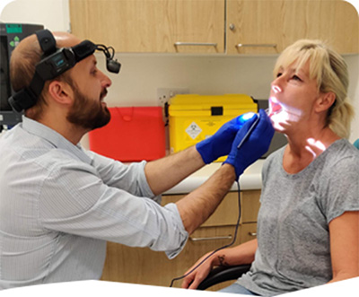
Red flags and first-line assessment
Urgent referral is indicated for progressive dysphagia, odynophagia, unexplained weight loss, persistent hoarseness or voice change, haemoptysis, a neck mass or lymphadenopathy, unilateral symptoms such as otalgia, and a history of heavy smoking or alcohol use [1]. These features take precedence over any reflux or muscle-tension explanation.
For patients without red flags, the history should establish the character of the sensation (constant or intermittent, worse with saliva or with food), its relationship to meals and posture, any heartburn or regurgitation, morning hoarseness, throat clearing, postnasal drip, snoring and sleep quality, and the occupational voice demand. Validated instruments help to quantify the burden and to monitor response. The Reflux Symptom Index (RSI) is widely used, though it does not capture all extra-oesophageal symptoms and its heartburn item is often excluded when assessing throat-predominant disease [2]. The EAT-10 is used where dysphagia is part of the picture.
Examination should include the oral cavity, oropharynx (tonsil size, tonsil stones, lingual tonsil bulk), neck palpation, and flexible nasendoscopy. Nasendoscopy allows assessment of the nasopharynx and postnasal space, the hypopharynx, the laryngeal inlet and the vocal folds, and is performed awake with the patient able to perform phonation and provocation manoeuvres. Findings are documented photographically where useful for follow-up.
Part 2 of the series follows on from Part 1. Watch it here, then read on for what you can do at home.
Self-Care at Home: What Patients Can Do
Many of the measures below are the foundation of management for reflux-related throat symptoms, and several are also recommended for globus without reflux. The evidence varies. Some measures are supported by randomised or controlled data, others by observational studies or consensus statements, and some are sensible common practice with limited direct evidence. The strength of each is stated so that patients and referring clinicians can judge them.
Eating patterns and timing
- Avoid large meals and finish eating at least two to three hours before lying down. A systematic review found that late evening meals increased the time the oesophagus was exposed to acid while lying down, compared with earlier meals [43]. Eating less in the evening is also supported by the European guideline on LPRD [51].
- Keep a food and drink diary for two to four weeks. Foods and drinks measurably change pepsin levels in saliva, and the pepsin concentration after meals predicted the response to treatment in one prospective cohort [53]. A diary helps identify individual triggers, which vary considerably between patients.
- Reduce refluxogenic foods and drinks. Caffeine, alcohol, carbonated drinks, chocolate, peppermint, fatty and fried foods, spicy food, and citrus and tomato-based foods are the most commonly identified triggers. In a crossover study of a low-fat, low-quick-release sugar, high-protein, alkaline, plant-based diet for six to twelve weeks, 74% of 50 patients with objectively confirmed LPR reported significant symptom improvement, and 54% continued to improve between six and twelve weeks [50]. This was uncontrolled, and the success rate was lower than the headline figure suggests, so diet should be viewed as an important adjunct rather than a stand-alone cure. The San Diego consensus also endorses lifestyle modification as appropriate for patients with coexisting oesophageal reflux symptoms [52]. Evidence: observational for the diet pattern; consensus support.
- Stay well hydrated through the day. Hydration keeps the throat moist and is part of the standard voice-hygiene advice [1]. Evidence: consensus and common practice.
Part 3 demonstrates exercises that can help the feeling of a lump in the throat. They fit alongside the self-care measures described here and below.
Body position and sleep
- Raise the head of the bed. In randomised comparisons summarised in a systematic review, elevating the head of the bed reduced the time spent with acid in the oesophagus when lying supine, from 21% to 15% [43]. A randomised crossover study showed that elevation on a foam wedge reduced the time distal oesophageal pH was below 4 compared with a flat position, while elevation on bed blocks showed a similar trend that was not statistically significant [45]. A systematic review of nocturnal reflux also concluded that bed-head elevation alleviates nocturnal heartburn and associated sleep disturbance [46]. Use bed blocks or a wedge placed under the mattress, not extra pillows, which bend the body at the waist and can increase pressure on the stomach. Evidence: randomised data for physiological effect; observational and small randomised data for symptoms.
- Sleep on the left side where comfortable. In a double-blind, sham-controlled randomised trial of an electronic sleep positional device, 44% of patients in the active group achieved at least a 50% reduction in nocturnal reflux score, compared with 24% in the sham group. The active device increased time spent sleeping on the left side [44]. An earlier small study of a positional sleep device also reported a significant reduction in nocturnal reflux symptoms over two weeks [47]. Left-side sleeping is supported by experimental work, and a positional device may help patients who find it difficult to maintain. Evidence: randomised (device); small studies for the wider benefit of position.
- Avoid slumping or lying down for at least one hour after eating. This is standard advice in the European and US reviews of lifestyle management [43][51]. Evidence: consensus.
- Avoid strenuous exercise immediately after a meal. Common practice, widely recommended. Evidence: consensus.
Weight and smoking
- If you are overweight, a structured weight reduction programme is one of the most effective reflux measures. In a prospective cohort of 332 overweight and obese adults, six months of structured weight loss led to complete resolution of GERD symptoms in 65% of participants, with the reduction in symptom score correlating with the amount of weight lost [48]. A population-based cohort (HUNT study) showed a dose-dependent association between weight loss and reduction in reflux symptoms, which was stronger among patients taking regular antireflux medication [49]. A systematic review also concluded that weight loss reduces oesophageal acid exposure [43]. Evidence: prospective cohort and population cohort; no randomised data in throat symptoms.
- Stop smoking. In a large prospective cohort, smoking cessation was associated with a substantial reduction in reflux symptoms, particularly in normal-weight individuals [43]. Smoking also irritates the upper airway and voice, and cessation benefits the throat and voice independently. Evidence: prospective cohort.
Voice and throat habits
- Stop clearing your throat repeatedly. Repeated throat clearing irritates the laryngeal lining and encourages further clearing, and avoiding habitual throat clearing is part of standard primary-care advice [1]. Replace it with a gentle swallow or a sip of water. Evidence: consensus; reflects the mechanism of the symptom.
- Rest your voice when it is hoarse, and avoid whispering. Whispering places more strain on the voice box than normal speech. Speech and voice therapy are effective for voice strain and muscle tension dysphonia, and a randomised trial of stretch-and-flow voice therapy showed a significant reduction in voice handicap compared with hygiene education alone [20]. Evidence: consensus for rest and avoidance of whispering; randomised data for voice therapy as a formal treatment.
- Use steam or a humidifier if the air is dry. Commonly advised for dry throat, particularly in heated or air-conditioned environments. Evidence: limited; common practice.
Stress and muscle tension
- Reduce stress where you can, and consider relaxation techniques, exercise such as walking or swimming, and gentle neck and shoulder stretches. Stress reduction is included in the European consensus statements for empirical treatment of LPRD [51], and a prospective study of lifestyle modification alone showed improvement in throat symptoms over two months, with improvement correlating with compliance [54]. Psychological therapy may help when anxiety is driving the symptom cycle [1]. Evidence: European consensus; prospective data for lifestyle programmes.
- Avoid tight clothing around the waist. Common practice, recommended in patient-facing reflux advice. Evidence: consensus.
Medicines available without prescription
- Alginate (Gaviscon Advance or equivalent). Alginates form a raft on top of the stomach contents and reduce exposure of the throat to reflux. European consensus recommends alginates or antacids for the empirical treatment of LPRD, addressing both acid and alkaline reflux events [51]. The San Diego consensus also supports alginate therapy when oesophageal reflux symptoms coexist [52]. A randomised trial in PPI-refractory reflux patients showed a modest benefit over placebo [11]. In a randomised trial of liquid alginate in LPRD, both alginate and placebo improved symptoms, so the specific benefit over placebo remains uncertain [10]. A pragmatic randomised trial in persistent throat symptoms is awaited [12]. Take the product as directed on the label, after meals and at bedtime, and continue for two weeks after symptoms settle before tapering. Evidence: consensus and randomised data, with the throat-specific question still open.
- Proton pump inhibitors. These are not recommended for throat symptoms alone. A large randomised, placebo-controlled trial found no benefit over placebo at 16 weeks or 12 months [7]. PPIs remain appropriate for patients with heartburn, regurgitation or dyspepsia, and the European consensus keeps PPIs for patients with acidic LPRD and GORD findings, for as short a period as possible (at least two months) with a planned reduction [51]. Do not stop a PPI abruptly if it has been prescribed for reflux disease, as rebound symptoms are common; discuss the plan with your GP.
When to seek help
If home measures have not improved symptoms after four to six weeks, if the sensation is getting worse, or if you develop any of the red flags listed above, please arrange a review. The San Diego consensus recommends upper endoscopy and ambulatory reflux monitoring when symptoms persist, when throat symptoms occur without oesophageal reflux symptoms, or when treatment needs escalation [52]. Please remember that a GP or specialist review is needed to make sure there is no other cause.
Laryngopharyngeal reflux (LPR)
Pathophysiology. The larynx and pharynx lack the robust protective mechanisms of the oesophagus. Mucosa in these regions is more susceptible to injury by pepsin and acid, and experimental work suggests that relatively few reflux events per week are sufficient to produce laryngeal damage [1]. Pepsin is stable and can persist in tissue and remain active on re-exposure, which may partly explain fluctuating symptoms [1]. This also helps to explain why LPR can occur without classic heartburn.
Diagnosis. There is no gold standard. Clinical scoring, most commonly the Reflux Finding Score (RFS), has been widely used, but it is subjective, shows substantial inter-observer variation, and has been reported to correlate poorly with pH evidence of reflux [3]. Objective testing, usually 24-hour multichannel intraluminal impedance with pH monitoring, is preferred where the diagnosis matters for treatment decisions. Recent consensus work supports objective testing to demonstrate LPR in patients with laryngopharyngeal symptoms, and highlights that pharyngeal reflux is frequently non-acidic or alkaline in nature [4]. Salivary pepsin has been proposed as a biomarker, with a recent meta-analysis reporting encouraging but heterogeneous accuracy [5]. Pharyngeal pH probes alone are not reliable for detecting all pharyngeal reflux events [6].
Treatment evidence. This is where the evidence has shifted most. A large multicentre, double-blind, placebo-controlled randomised trial of lansoprazole 30 mg twice daily for 16 weeks in 346 patients with persistent throat symptoms found no benefit over placebo at 16 weeks (estimated difference 1.9 RSI points in favour of placebo) or at 12 months [7]. A systematic review of earlier PPI trials had likewise found that randomised trials did not show a clear advantage of PPIs over placebo [8]. Consequently, empirical PPI therapy for throat symptoms in the absence of heartburn or other typical reflux symptoms is not supported by current evidence.
Alginate therapy is supported mechanistically. Because the predominant reflux events in the pharynx may be weakly acid or alkaline, and because alginate forms a raft that limits exposure of the upper aerodigestive tract to refluxate, alginates are increasingly preferred over acid suppression in this setting, and a recent review concluded that the evidence increasingly supports alginate and magaldrate over PPIs for pharyngeal reflux [4][9]. In a randomised trial of an alginate suspension in LPRD patients, both alginate and placebo significantly reduced symptoms and reflux numbers, but the alginate was not superior to placebo, and the authors emphasised a large placebo response and the importance of lifestyle modification [10]. Trial data in reflux patients with inadequate response to PPI do show a modest benefit of Gaviscon Advance over placebo for reflux symptoms [11]. A pragmatic multicentre randomised trial of liquid alginate in persistent throat symptoms (ISRCTN13949559) has been registered and is designed to deliver definitive evidence at eight weeks [12]. Until that is available, the honest position is that alginates are a rational and low-risk first-line option, supported by mechanism and by some clinical data, but not yet proven superior to placebo in throat-predominant disease.
Practical regimen. In clinic we advise lifestyle and dietary measures as the foundation: reducing caffeine, alcohol, carbonated drinks, chocolate, peppermint, fatty foods and very salty or spicy foods; finishing meals at least two to three hours before lying down; elevating the head of the bed; and avoiding slumping or bending for an hour after eating. An alginate (typically a 10 to 20 mL dose after meals and at bedtime, per the product label) is commenced for a defined period, with a planned taper after symptoms settle. Patients should be reminded that recurrence is common and that early re-treatment at the first sign of relapse is more effective than waiting for established symptoms.
Where PPIs remain indicated. PPIs remain appropriate for patients with heartburn, regurgitation or dyspepsia, for confirmed erosive disease, and for eosinophilic oesophagitis, where PPI response is part of the diagnostic and therapeutic pathway (see below). Duration should be limited and reviewed, given the rebound phenomenon and the ongoing discussion about long-term adverse effects.
Cervical inlet patch (cervical heterotopic gastric mucosa)
Cervical inlet patches (CIPs) are islands of heterotopic gastric mucosa in the cervical oesophagus, typically at or just below the upper oesophageal sphincter. They are thought to be congenital ectopic gastric mucosa, although metaplastic origin has also been proposed [13]. Prevalence is reported between 1% and 21% on endoscopy, and autopsy series suggest that detection is substantially under-reported [14]. Detection depends on deliberate inspection of the proximal oesophagus, and it has been suggested that awareness alone can increase endoscopic detection markedly [14].
Diagnosis. Patches appear as salmon-coloured, velvety mucosa with well-defined margins, contrasting with the grey-white stratified squamous epithelium of the normal oesophagus. Endoscopic appearance should be confirmed by biopsy demonstrating gastric-type mucosa adjacent to normal oesophageal mucosa, and the proximal oesophagus should be examined carefully where globus is the presenting symptom. Barium swallow may show indentations or rim-like shadows at the margins of the patch, but its overall value in globus is limited [1].
Symptoms and association. Patches are most often asymptomatic. When symptomatic, they can cause globus, sore throat, cough and, in some cases, dysphagia and peptic stricture due to acid and pepsin secretion [14]. Symptom relief after ablation has been reported in several series [15][16][17], and in a sham-controlled randomised trial of argon plasma coagulation, 9 of 11 treated patients reported improvement compared with none of 10 controls at three months, with long-term benefit in 13 of 17 treated patients after a median follow-up of 17 months [15]. These trials are small, and the mechanism of benefit is not established. Some clinicians have raised the possibility that stretching of the cricopharyngeus during endoscopy contributes to the effect, which has not been excluded [15].
Treatment. Asymptomatic patches require no treatment. Symptomatic patches are treated with a course of PPI, typically for four to six weeks, to assess response, followed by endoscopic ablation if symptoms persist. Argon plasma coagulation (APC) and through-the-scope radiofrequency ablation (RFA) have both been used. In a prospective pilot study of through-the-scope RFA in ten patients, 80% achieved complete endoscopic and histological resolution after a median of two sessions, with significant improvement in globus, sore throat and cough at one-year follow-up [16]. In a retrospective cohort of 45 patients treated with APC, 31% reported a very strong improvement and 24% a strong improvement, with complete GIP eradication after one session in 56% [17]. Ablation is repeated where residual patch is present. Strictures are a recognised complication of ablation of large or circumferential patches, and treatment is therefore staged, ablating one hemi-circumference at a time where the patch is large [18].
Our position. Ablation is reasonable for patients with clearly documented, symptomatic patches in whom PPI and conservative management have failed, after a careful risk-benefit discussion. Ongoing randomised work is awaited to determine whether symptom improvement is attributable to ablation of the patch rather than to dilatation of the cricopharyngeal region, and to define selection criteria. Patients should be informed that the evidence is currently limited to small series and one small sham-controlled trial.
Muscle tension dysphonia and laryngeal muscle tension
Muscle tension dysphonia (MTD) is a functional voice disorder characterised by excessive and asymmetrical laryngeal muscle activity, which causes laryngeal hyperfunction, a strained or hoarse voice, and sensation of tightness, lump or effort in the throat [19]. It can be secondary to laryngitis, reflux, an upper respiratory infection or voice overuse, and is perpetuated by compensatory strain. The condition is known by several names, including functional dysphonia, vocal hyperfunction, muscle misuse dysphonia and muscle imbalance dysphonia.
Assessment. Diagnosis is clinical, supported by laryngoscopic evidence of supraglottic constriction or hyperadduction, and by perceptual voice assessment and acoustic measures such as the Voice Handicap Index and cepstral peak prominence. Concurrent laryngeal pathology, including reflux-related changes, must be excluded before attributing symptoms to MTD alone.
Treatment. Voice therapy by a speech and language therapist specialising in voice is the first-line treatment and is effective in many patients. A randomised controlled trial of stretch-and-flow voice therapy in primary MTD showed a significantly greater reduction in Voice Handicap Index compared with vocal hygiene education alone, with a large effect size, although the sample was small [20]. A retrospective audit of 68 patients suggested that specific techniques, including optimal phonation tasks and negative practice, contributed measurable improvement, supporting the use of defined therapy components [21]. Laryngeal manual therapy has shown acoustic benefit in a recent systematic review and meta-analysis, although the authors noted that high-quality trials are still needed [22]. Multimodal physiotherapy incorporating myofascial release has been reported to add benefit to voice therapy in patients with cervicalgia and functional laryngeal symptoms, including globus [23].
Injection laryngoplasty is occasionally considered as an adjunct in patients who have had a suboptimal response to voice therapy, and in one retrospective series of 37 patients, voice-related quality of life improved, with the greatest acoustic change in those with glottal insufficiency [24]. This is a specialist option and is not appropriate as a first-line treatment.
Lingual and palatine tonsil hypertrophy
Anatomy and symptoms. Hypertrophy of the palatine tonsils can produce a sensation of a lump through contact between enlarged tonsils, and infected or stone-laden tonsils can cause a persistent foreign-body sensation disproportionate to their size. Lingual tonsil hypertrophy can cause a globus-like sensation, dysphagia, airway obstruction and, importantly, mechanical contact with the epiglottis, which may alter epiglottic shape and produce a sensation of fullness [25].
Assessment. Palatine tonsil size is graded clinically. Lingual tonsil size is assessed by direct inspection and flexible endoscopy, and the contribution of the lingual tonsil to upper airway obstruction is best assessed during sleep endoscopy where sleep-disordered breathing is suspected. In patients with persistent symptoms and no other explanation, a tonsillar contribution should be considered alongside reflux and muscle tension.
Treatment. Palatine tonsillectomy is considered where tonsil size, recurrent infection, tonsil stones or airway obstruction are clearly contributing, subject to standard criteria. For lingual tonsil hypertrophy, endoscopic-assisted coblation or radiofrequency lingual tonsillectomy is an established technique. In a case series of 26 children with persistent obstructive sleep apnoea after adenotonsillectomy and lingual tonsil hypertrophy, the respiratory disturbance index fell from a mean of 14.7 to 8.1 following endoscopic-assisted coblation [26]. Surgeons have described advantages of coblation for lingual tonsillectomy, including improved visualisation, less bleeding and less postoperative pain than laser or monopolar techniques, although these are largely supported by case series and surgical technique papers [27][28][29]. For palatine tonsillectomy, a Cochrane review found that coblation may cause less pain on the first postoperative day, but that the difference is small and by day seven there appears to be little or no difference. Primary bleeding risk was similar, and secondary bleeding risk may be slightly higher, although the evidence is of low quality [30]. More information is on our Tonsils & Adenoids page, with consent information for tonsillectomy and lingual tonsil surgery.
"After a lot of research, I chose Vik Veer as my surgeon to perform a tonsillectomy. From my initial consultation, I felt at ease and his consultation was thorough and extremely informative. The surgery went well and I would have no hesitation in recommending Mr Veer." Google Review, 5 stars — Melanie Treadwell
Eosinophilic oesophagitis
Eosinophilic oesophagitis (EoE) is a chronic, immune-mediated disease of the oesophagus, diagnosed on the basis of symptoms of oesophageal dysfunction and an eosinophil-predominant infiltrate, conventionally at least 15 eosinophils per high-power field on biopsy [31][32]. Its incidence and prevalence are rising, and it is increasingly encountered in primary care and ENT settings as well as in gastroenterology [31].
The symptom overlap with reflux is considerable, including retrosternal burning and regurgitation. Features that favour EoE include onset of symptoms during or shortly after the first few bites of a meal, prominent dysphagia, and food impaction. Endoscopic features include linear furrows, white plaques, concentric rings, and a narrow calibre oesophagus, although the mucosa can appear normal in a substantial proportion of patients [31][33].
The diagnostic pathway has changed. Earlier guidance required a PPI trial before EoE could be diagnosed. The international consensus criteria (AGREE) removed the PPI-trial requirement and recognise PPIs as a treatment for oesophageal eosinophilia that may be due to EoE, rather than as a diagnostic test [32]. A PPI-responsive subgroup has been described, which is clinically and histologically indistinguishable from EoE in many respects [34]. In an open-label study of double-dose omeprazole for eight weeks in patients with eosinophilic infiltration in the EoE range, more than half showed clinical improvement, and about 37% achieved both clinical and histological remission [35].
Treatment is multimodal: PPIs, topical corticosteroids, empiric elimination diets, and oesophageal dilatation for strictures [31][36]. Management in an ENT setting should be co-ordinated with gastroenterology, and biopsies should be taken from the oesophagus when EoE is suspected, with a low threshold in patients with food impaction, dysphagia, or globus with reflux-type symptoms in younger patients.
Pharyngeal pouch (Zenker’s diverticulum)
A pharyngeal pouch is a pulsion diverticulum arising through Killian’s dehiscence, just above the cricopharyngeus muscle, in the posterior wall of the hypopharynx [37]. Small pouches can be asymptomatic or cause occasional sensation of food sticking in the lower throat. Larger pouches collect food and fluid, causing dysphagia (the commonest symptom), regurgitation of undigested food, halitosis, cough and aspiration. Severe disease can cause weight loss and recurrent chest infection.
Diagnosis. A gastrografin swallow is the most useful initial test, giving a rapid, safe assessment of pouch size and position. Barium studies, endoscopy and high-resolution manometry may be used to complete the assessment, particularly to exclude other pathology. The EAT-10 questionnaire is used to quantify swallowing burden, with a score of 3 or more considered abnormal and a score of 15 or more associated with higher risk of aspiration [38].
Treatment. Symptomatic pouches are treated surgically, and the goal is division of the cricopharyngeal muscle to relieve outflow obstruction, with or without pouch resection. Endoscopic approaches are preferred where anatomy permits, and open cervical surgery is reserved for pouches that cannot be reached endoscopically. Rigid endoscopic stapling offers symptom relief in around 90% of cases, with lower morbidity and short hospital stays of one to two days, but anatomical limitations restrict its use [39]. Endoscopic CO2 laser cricopharyngeal myotomy has comparable safety and efficacy in a retrospective cohort of 108 patients, with no significant difference in persistence or recurrence of symptoms between laser and stapling, although hospital stay was longer with laser in that series [40]. Z-POEM (peroral endoscopic myotomy) has emerged as a minimally invasive option, with clinical success reported between 85% and 93% and major complications in roughly 5%, although long-term recurrence data are still emerging [37]. In a comparative cohort of 424 patients, recurrence was higher after several endoscopic techniques, including rigid stapling and flexible endoscopic approaches, and was lowest after open and endoscopic laser approaches [41]. A systematic review found that bleeding and recurrence were more likely after flexible endoscopic techniques, whereas rigid approaches resulted in fewer revision surgeries [42].
Perforation is the principal serious complication of endoscopic pouch surgery. Perforations are uncommon but can be serious, and they require antibiotics, nil by mouth with feeding via a tube, and inpatient observation, with some requiring further surgery [42].
Other contributors
Postnasal drip and upper airway inflammation. Allergic rhinitis and chronic rhinosinusitis can produce postnasal drip and local sensitivity that manifests as globus. Nasal examination and treatment of nasal disease are part of the assessment. See our guide to postnasal drip.
Cervical spine abnormalities. Osteophytes and postural abnormalities can cause referred throat sensation, and are identified on imaging where clinically indicated [1].
Psychological factors. Anxiety and psychogenic factors contribute to, and are amplified by, globus. Reassurance and explanation are essential components of care, and cognitive behavioural approaches may help in patients with significant distress [1].
Sleep-disordered breathing. Upper airway obstruction during sleep, and night-time reflux driven by negative intrathoracic pressure, can worsen throat symptoms. Where morning symptoms predominate or snoring is significant, sleep assessment should be considered. See snoring and obstructive sleep apnoea and our sleep study service.
Investigations in summary
- Flexible nasendoscopy in clinic: first-line, with assessment of the hypopharynx, laryngeal inlet and vocal folds.
- Barium swallow: useful for pouch, large patches, or structural causes of dysphagia; limited value for routine exclusion of malignancy in globus [1].
- Upper GI endoscopy with examination of the proximal oesophagus: indicated for dysphagia, red flags, suspected inlet patch or EoE.
- Objective reflux testing (multichannel intraluminal impedance with pH): where the diagnosis determines treatment, and where empirical treatment has failed.
- High-resolution oesophageal manometry: where dysphagia suggests motility disorder or before surgery for pouch or reflux.
- MRI or CT of the neck: reserved for high-risk patients or where an abnormality is identified on examination.
What to Expect at a Consultation
Professor Veer will take a detailed history of your symptoms and perform a thorough examination including flexible nasendoscopy where indicated. This is a brief, well-tolerated procedure in which a thin, flexible camera is passed through the nostril to view the back of the nose, throat, and vocal cords in real time. It takes approximately two minutes and provides immediate information that significantly aids diagnosis.
Following your consultation you will receive a clear explanation of findings, a diagnosis where possible, and a structured management plan. The same assessment applies to other throat symptoms such as a persistent sore throat, a hoarse voice or difficulty swallowing.
"I just wanted to express my heartfelt gratitude for the seamless care I received during my operation. A huge thank you to Dr Veer for his thorough and professional approach, and to Kerry for her flawless support in keeping everything organised. Your kindness, dedication, and expertise truly made a difference." Google Review, 5 stars — Ama
Summary
Globus is a symptom with several contributors that often coexist. A structured approach that excludes red flags, examines the whole upper aerodigestive tract, and considers reflux, inlet patches, tonsils, muscle tension, eosinophilic oesophagitis and pharyngeal pouch in turn, leads to better outcomes than treating a single presumed cause. Current evidence does not support PPIs for throat symptoms alone, alginate therapy is a rational first-line option with evidence still being developed, and surgical or endoscopic options should be reserved for patients with well-defined pathology who have failed conservative measures.
Frequently Asked Questions
Globus pharyngeus is a persistent sensation of a lump, tightness or foreign body in the throat when there is no structural obstruction and no true difficulty swallowing. It is common: an estimated 45% of people experience it at some point, and it accounts for roughly 4% of ENT referrals. It is best understood as a symptom with several possible contributors rather than a single diagnosis.
In most people the cause is benign, but globus can overlap with malignancy, neuromuscular disease and oesophageal disease, so the first step is always to exclude serious causes. Urgent assessment is needed for progressive difficulty swallowing, painful swallowing, unexplained weight loss, persistent hoarseness or voice change, coughing up blood, a lump in the neck, one-sided symptoms such as ear pain, or a history of heavy smoking or alcohol use.
Four groups account for most cases: laryngopharyngeal reflux (silent reflux), upper oesophageal and cervical inlet pathology such as a cervical inlet patch, laryngeal muscle tension, and structural factors in the upper aerodigestive tract such as enlarged lingual or palatine tonsils or a pharyngeal pouch. Less commonly, eosinophilic oesophagitis causes a similar picture. Postnasal drip, cervical spine problems, anxiety and sleep-disordered breathing can also contribute.
Not for throat symptoms on their own. A large randomised, placebo-controlled trial of lansoprazole in 346 patients with persistent throat symptoms found no benefit over placebo at 16 weeks or at 12 months. PPIs remain appropriate if you also have heartburn, regurgitation or indigestion. An alginate such as Gaviscon Advance is a rational, low-risk first-line option, although it has not yet been proven superior to placebo in throat-predominant disease.
Measures with supporting evidence include finishing meals two to three hours before lying down, reducing caffeine, alcohol, fizzy drinks, chocolate, peppermint and fatty or spicy food, raising the head of the bed on blocks or a wedge, sleeping on the left side where comfortable, losing weight if you are overweight, stopping smoking, avoiding repeated throat clearing, staying hydrated and reducing stress. If symptoms have not improved after four to six weeks, or any red-flag symptom develops, you should be reviewed.
Assessment begins with a detailed history and examination of the mouth, tonsils and neck, followed by flexible nasendoscopy, in which a thin camera is passed through the nose to view the back of the nose, throat, voice box and vocal cords. It takes about two minutes and is performed awake. Depending on the findings, further tests may include a barium swallow, upper gastrointestinal endoscopy, 24-hour impedance-pH reflux monitoring, oesophageal manometry or, rarely, MRI or CT of the neck.
Only for well-defined pathology in patients who have not improved with conservative measures. Examples are ablation of a symptomatic cervical inlet patch, tonsillectomy or coblation lingual tonsillectomy when tonsil tissue is clearly contributing, and surgery for a symptomatic pharyngeal pouch. The evidence for some of these options is limited to small series, so the risks and benefits are discussed in detail before any decision.
Yes. Upper airway obstruction during sleep, and night-time reflux driven by negative pressure in the chest, can worsen throat symptoms. If your symptoms are worse in the morning or you snore significantly, a sleep assessment should be considered alongside throat treatment.
References
- Muddaiah A, Watson A, Chopra R. Globus: a practical guide for general practice. Br J Gen Pract. 2025;75(754):237-238. doi:10.3399/bjgp25X741573
- Lechien JR, et al. Instruments evaluating the clinical findings of laryngopharyngeal reflux: a systematic review. Laryngoscope. 2019;129(3):720-736.
- Belafsky PC, Postma GN, Koufman JA. The validity and reliability of the reflux finding score (RFS). Laryngoscope. 2001;111(8):1313-1317.
- Lechien JR, et al. Evaluation and management of laryngopharyngeal reflux disease: an updated state of the art review. Otolaryngol Head Neck Surg. 2026. doi:10.1002/ohn.70413
- Wang J, Zhao Y, Ren J, et al. Pepsin in saliva as a diagnostic biomarker in laryngopharyngeal reflux: a meta-analysis. Eur Arch Otorhinolaryngol. 2018;275(3):671-678.
- Desjardin M, Roman S, des Varannes SB, et al. Pharyngeal pH alone is not reliable for the detection of pharyngeal reflux events: a study with oesophageal and pharyngeal pH-impedance monitoring. United European Gastroenterol J. 2013;1(6):438-444.
- O’Hara J, et al. Use of proton pump inhibitors to treat persistent throat symptoms: multicentre, double blind, randomised, placebo controlled trial. BMJ. 2021. doi:10.1136/bmj.m4903
- Karkos PD, et al. Empiric treatment of laryngopharyngeal reflux with proton pump inhibitors: a systematic review. Laryngoscope. 2006.
- Lechien JR, et al. Evaluation and management of laryngopharyngeal reflux disease: state of the art review. Otolaryngol Head Neck Surg. 2019;160(5):762-782.
- Tseng W-H, et al. Double-blind, placebo-controlled study with alginate suspension for laryngopharyngeal reflux disease. Laryngoscope. 2018;128:2252-2260.
- Reimer C, et al. Randomised clinical trial: alginate (Gaviscon Advance) vs. placebo as add-on therapy in reflux patients with inadequate response to a once daily proton pump inhibitor. Aliment Pharmacol Ther. 2016. doi:10.1111/apt.13567
- Morton M, et al. Trial of alginates in throat symptoms: protocol for a pragmatic, multicentre, placebo controlled, double blind, parallel, randomised controlled trial of liquid alginate (Gaviscon Advance). BMJ Open. 2025. doi:10.1136/bmjopen-2024-087729
- Meining A, et al. Argon plasma ablation of gastric inlet patches in the cervical esophagus may alleviate globus sensation: a pilot trial. Endoscopy. 2006;38:566-570.
- Yang YH, et al. An obscure case of dysphagia. Am J Gastroenterol. 2024. (Conference abstract.)
- Bajbouj M, et al. Argon plasma coagulation of cervical heterotopic gastric mucosa as an alternative treatment for globus sensations. Gastroenterology. 2009;137:440-444.
- Dunn J, et al. Radiofrequency ablation of symptomatic cervical inlet patch using a through-the-scope device: a pilot study. Gastrointest Endosc. 2016.
- Brechmann T, et al. Argon plasma coagulation of gastric inlet patches of the cervical esophagus relieves vocal and respiratory symptoms in selected patients. Dig Dis Sci. 2022. doi:10.1007/s10620-022-07745-9
- Yang YH, et al. An obscure case of dysphagia (stricture following staged ablation of a circumferential patch). Am J Gastroenterol. 2024. (Conference abstract.)
- Tomlinson CA, et al. Manual therapy and exercise to improve outcomes in patients with muscle tension dysphonia: a case series. Phys Ther. 2014.
- Watts CR, et al. A randomized controlled trial of stretch-and-flow voice therapy for muscle tension dysphonia. Laryngoscope. 2015.
- Madill C, et al. Active ingredients of voice therapy for muscle tension voice disorders: a retrospective data audit. J Clin Med. 2021. doi:10.3390/jcm10184135
- Latoszek BBV, et al. The efficacy of manual circumlaryngeal therapy for muscle tension dysphonia: a systematic review and meta-analysis. Laryngoscope. 2024;134:18-26.
- Gillespie M, et al. Myofascial release therapy augments the effect of voice therapy in patients with cervicalgia. Laryngoscope. 2025.
- Novakovic D, et al. Injection laryngoplasty as adjunct treatment method for muscle tension dysphonia: preliminary findings. Laryngoscope. 2020;130:980-985.
- Epiglottic displacement by lingual or palatine tonsil hypertrophy: described in the clinical literature.
- Lin A, et al. Persistent pediatric obstructive sleep apnea and lingual tonsillectomy. Otolaryngol Head Neck Surg. 2009. doi:10.1016/j.otohns.2009.03.011
- Maturo SC, Mair EA. Coblation lingual tonsillectomy. Otolaryngol Head Neck Surg. 2006. doi:10.1016/j.otohns.2006.04.015
- Robinson S, et al. Lingual tonsillectomy using bipolar radiofrequency plasma excision. Otolaryngol Head Neck Surg. 2006. doi:10.1016/j.otohns.2005.10.021
- Rotenberg B, et al. Endoscopic-assisted radiofrequency lingual tonsillectomy. Laryngoscope. 2011. doi:10.1002/lary.21730
- Pynnonen M, et al. Coblation versus other surgical techniques for tonsillectomy. Cochrane Database Syst Rev. 2017;CD004619. doi:10.1002/14651858.CD004619.pub3
- Dellon ES, et al. ACG clinical guideline: diagnosis and management of eosinophilic esophagitis. Am J Gastroenterol. 2025. doi:10.14309/ajg.0000000000003194
- Dellon ES, et al. Updated international consensus diagnostic criteria for eosinophilic esophagitis: proceedings of the AGREE conference. Gastroenterology. 2018. doi:10.1053/j.gastro.2018.07.009
- Gonsalves N, et al. Diagnosis and treatment of eosinophilic esophagitis. J Allergy Clin Immunol. 2020. doi:10.1016/j.jaci.2019.11.011
- Molina-Infante J, et al. Proton pump inhibitor-responsive oesophageal eosinophilia: an entity challenging current diagnostic criteria for eosinophilic oesophagitis. Gut. 2015. doi:10.1136/gutjnl-2015-310991
- Vázquez-Elizondo G, et al. The outcome of patients with oesophageal eosinophilic infiltration after an eight-week trial of a proton pump inhibitor. Aliment Pharmacol Ther. 2013. doi:10.1111/apt.12513
- Dellon ES, et al. ACG clinical guideline: evidence-based approach to the diagnosis and management of esophageal eosinophilia and eosinophilic esophagitis. Am J Gastroenterol. 2013. doi:10.1038/ajg.2013.71
- Ramamurthy S, et al. Management strategies for Zenker’s diverticulum: a comprehensive review. J Clin Med. 2025. doi:10.3390/jcm14176141
- Belafsky PC, Mouadeb DA, Rees CJ, et al. Validity and reliability of the Eating Assessment Tool (EAT-10). Ann Otol Rhinol Laryngol. 2008;117(12):919-924.
- Philippsen LP, et al. Endoscopic stapled diverticulotomy: treatment of choice for Zenker’s diverticulum. Laryngoscope. 2000;110(8). doi:10.1097/00005537-200008000-00011
- Dhar P, et al. Endoscopic laser pharyngeal pouch surgery: is it safe? Br J Surg. 2022. doi:10.1093/bjs/znac269.194 (Conference abstract.)
- Wallerius KP, et al. Comparing open versus rigid endoscopic and flexible endoscopic techniques for the treatment of Zenker’s diverticulum. Otolaryngol Head Neck Surg. 2023. doi:10.1002/ohn.318
- Crawley B, et al. Adverse events after rigid and flexible endoscopic repair of Zenker’s diverticula: a systematic review and meta-analysis. Otolaryngol Head Neck Surg. 2019. doi:10.1177/0194599819839991
- Ness-Jensen E, Hveem K, El-Serag H, et al. Lifestyle intervention in gastroesophageal reflux disease. Clin Gastroenterol Hepatol. 2016;14(2):175-182. doi:10.1016/j.cgh.2015.04.176
- Schuitenmaker JM, et al. Sleep positional therapy for nocturnal gastroesophageal reflux: a double-blind, randomized, sham-controlled trial. Clin Gastroenterol Hepatol. 2022. doi:10.1016/j.cgh.2022.02.058
- Hamilton JW, Boisen RJ, Yamamoto DT, et al. Sleeping on a wedge diminishes exposure of the esophagus to refluxed acid. Dig Dis Sci. 1988;33(5):518-522. doi:10.1007/bf01798350
- Gerson L, et al. A systematic review of the definitions, prevalence, and response to treatment of nocturnal gastroesophageal reflux disease. Clin Gastroenterol Hepatol. 2009. doi:10.1016/j.cgh.2008.11.021
- Allampati S, et al. Use of a sleep positioning device significantly improves nocturnal gastroesophageal reflux symptoms. Am J Gastroenterol. 2014. (Conference abstract; small pilot.)
- Singh M, et al. Weight loss can lead to resolution of gastroesophageal reflux disease symptoms: a prospective intervention trial. Obesity. 2012. doi:10.1002/oby.20279
- Ness-Jensen E, et al. Weight loss and reduction in gastroesophageal reflux: a prospective population-based cohort study (the HUNT study). Am J Gastroenterol. 2013. doi:10.1038/ajg.2012.466
- Lechien JR, et al. Is diet sufficient as laryngopharyngeal reflux treatment? A cross-over observational study. Laryngoscope. 2022;132:1916-1923. doi:10.1002/lary.29890
- Lechien JR, et al. European clinical practice guideline: managing and treating laryngopharyngeal reflux disease. Eur Arch Otorhinolaryngol. 2024. doi:10.1007/s00405-024-09181-z
- Yadlapati RH, et al. The San Diego consensus for laryngopharyngeal symptoms and laryngopharyngeal reflux disease. Am J Gastroenterol. 2025. doi:10.14309/ajg.0000000000003482
- Lechien JR, et al. Saliva pepsin concentration of laryngopharyngeal reflux patients is influenced by meals consumed before the samples. Laryngoscope. 2021;131:350-359. doi:10.1002/lary.28756
- Steward D, et al. Proton pump inhibitor therapy for chronic laryngo-pharyngitis: a randomized placebo-control trial. Otolaryngol Head Neck Surg. 2004. doi:10.1016/j.otohns.2004.03.037
This page was created on 29th October 2026 and is for general information. It does not replace assessment by a doctor. If you have any of the red-flag symptoms described above, please seek medical review promptly.